
Forschung
Overview
Our current main research interest lies in the development and improvement of dosimetry for radionuclide therapy with focus on oncological treatments. The aim is to provide a robust estimate of both, the radiation exposure to normal organs and the radiation damage to tumorous tissue, after administration of a radioactive substance (radiopharmaceutical). In this way, the risk of organ toxicities shall be reduced, while the probability to stop or at least control tumor growth shall be maximized (therapeutic window). Furthermore, dosimetry is a vital aspect to investigate mechanisms relevant for tumor response.
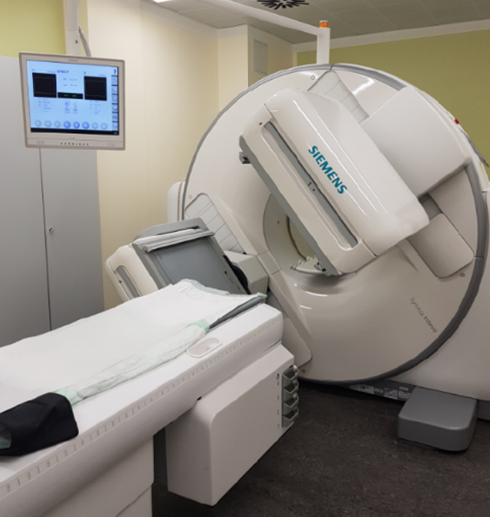 Quantitative molecular imaging via SPECT (Single-Photon-Emission-Computed-Tomography) or PET (Positron-Emission-Tomography) allows for a direct measure of the radionuclide distribution within the patient. Quantitative imaging is still a research field of interest, as it requires specific effort during the patient examination itself (e. g. patient positioning) and with regard to image reconstruction to correct for physical image degradation (e. g. detector response modelling). The accuracy of image reconstruction can be validated against Monte Carlo simulations of the desired imaging system. In addition to PET or SPECT, the patient anatomy can be imaged via accompanying Computed Tomography (CT) (hybrid SPECT/CT (s. Abbildung) or PET/CT). Automatic or semi-automatic image segmentation methods (e. g. kmeans clustering) are required to provide a fast, robust and user-independent detection of both, risk organs and tumorous tissue in those acquired images.
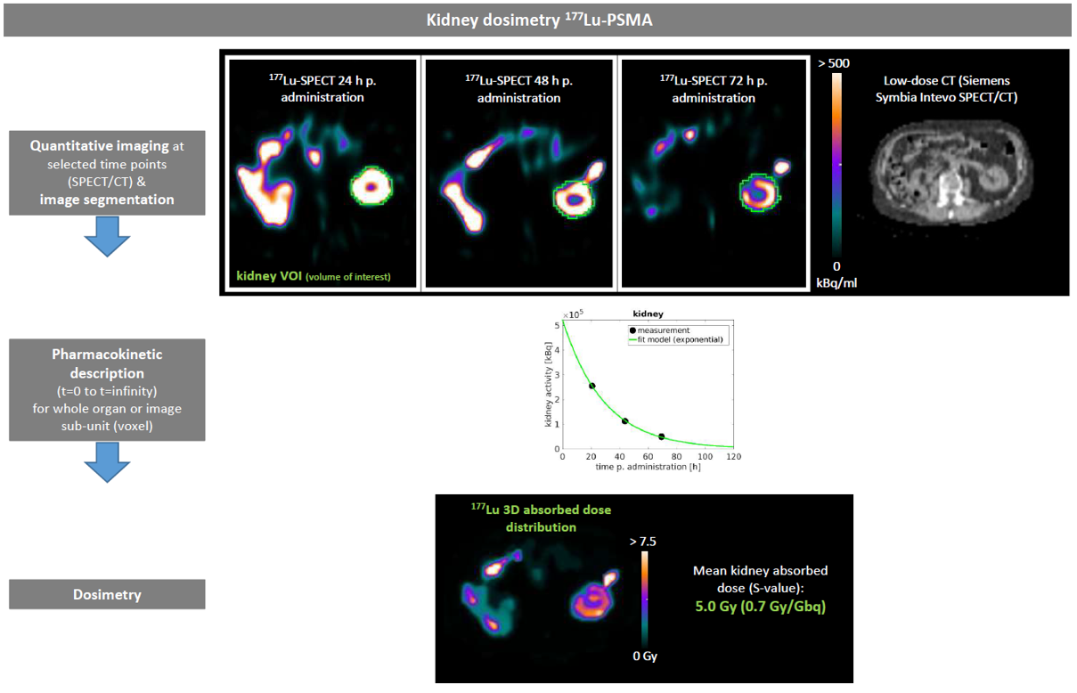
Dosimetry ideally requires the complete knowledge of the time course of radionuclide accumulation within all relevant tissues from radiopharmaceutical injection until the last radioactive decay. However, for a clinical surrounding, measuring the radionuclide distribution within the patient is only possible for a few selected time points (usually 3-4). Thus, appropriate mathematical curve models have to be fitted to these few data points to estimate the complete time course (time-activity-curve (TAC)). In doing so, time points of patient examination, mathematical fit model, and the pharmaceutical-specific accumulation have to be carefully synchronized (pharmacokinetic description).
Using the determined time-activity-curves, the physically absorbed dose of risk organs and lesions can be derived, as the physical properties of the employed radionuclide are known. More advanced 3D dosimetry techniques (Monte Carlo simulations) can model all relevant physical interactions underlying the radiation exposure within the patient after radionuclide administration for each single image sub-unit. However, Monte Carlo simulations are time-consuming and their routine clinical application is limited, compared to, e. g., less computational demanding but also potentially less accurate absorbed dose conversion factors (S-values). Thus, appropriate dosimetry methods have to be selected according to the required accuracy and clinical feasibility.
To summarize, dosimetry for radionuclide therapy is a demanding multi-step process and its integration into clinical routine is strongly limited by clinical capabilities. Thus, we work in close connection with physicians, technicians, and chemists to develop standards as accurate as possible in agreement with clinical feasibility and patient comfort. To provide dosimetry solutions with a high degree of automatization (e. g. automatic image segmentation) is mandatory to allow for an application in daily clinical routine.
Research topics
Dosimetry is vital to assess the radiation exposure to healthy tissues to avoid organ toxicities and to verify an absorbed dose to malignant tissues that is sufficient to achieve at least tumor control. Despite its outstanding role, dosimetry for radionuclide therapy is still an open research field and its routine application is still hardly integrated into clinical routine. One the one hand, the complete dosimetry chain for radionuclide therapy is complex, time-consuming, and so far lacks standardization. Furthermore, the uncertainty of absorbed dose values is still high, in particular for tumors, which significantly complicates to establish relationships between dosimetry and the understanding of tumor response.
Thus, our focus is to improve the accuracy, robustness and efficiency of dosimetry for radionuclide therapy to allow for both, a more frequent integration of dosimetry into clinical routine and a more clinical significance of dosimetry. In this sense, we are working on:
- SPECT/PET imaging for radionuclide therapy,
- Quantitative SPECT reconstruction,
- Monte Carlo simulations for SPECT imaging and absorbed dose modelling,
- Pharmacokinetic description of radiopharmaceuticals,
- Patient-specific 3D dosimetry techniques,
- Image segmentation.
There are permanently projects for Bachelor or Master projects or student assistants available on these topics. Larger work packages can also be offered as PhD thesis.
What we offer:
- Working in close connection to the clinical environment within a highly interdisciplinary team
- Highly diversifies tasks that can be focused according to personal interests and strengths
- Possibility to gain insight into future clinical working areas (e. g. Medical Physics Expert)
- Possibility to present research findings at national and international conferences
- Possibility to contribute to industry collaborations
In case of interest to participate in our research group, please send your request and CV to nuklearmedizin.physik@med.uni-muenchen.de. An existing knowledge on programming is not specifically required, however, in case of interest projects with stronger focus on programming skills can be offered (e .g. image reconstruction or automatic image segmentation).
Clinical background
The goal of radionuclide therapy is to achieve a highly selective irradiation of tumor cells via short-range radioactive decay products (electrons or alpha particles). The selective accumulation of the radioactive substance within the tumor tissue can be achieved, 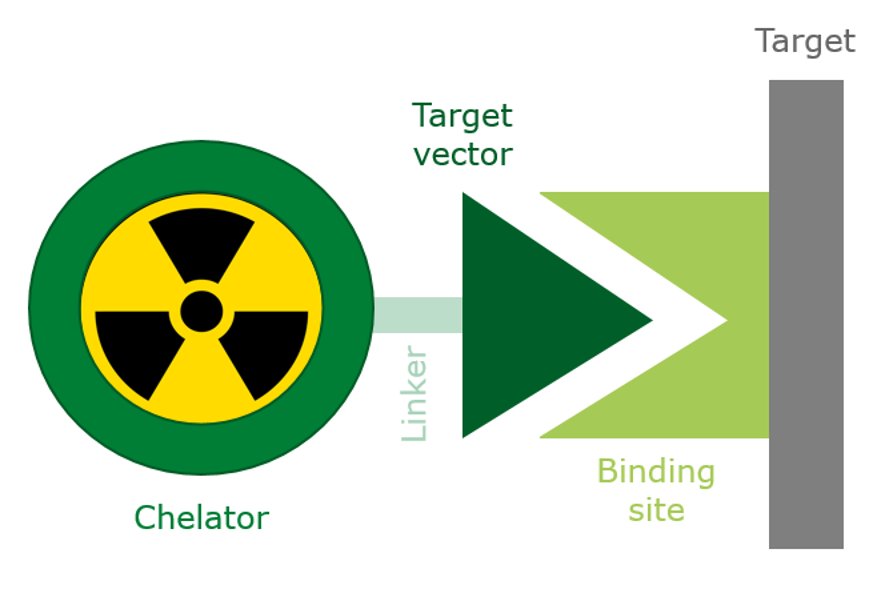 e.g., by metabolic trapping mechanisms or by localized administration (e.g. Iodine-131 for malignant or benign thyroid disease or Selective Internal Radiotherapy (SIRT) using Yttrium-90 for treatment of liver metastases). Many available radiopharmaceuticals employ so-called carrier molecules, which have the ability to target binding sites that are over- or specifically expressed by cancer cells. The design of such carrier-based pharmaceuticals is guided by the Theranostic concept. The Theranostic concept refers to the aim to label a specific carrier molecule either with therapeutic beta-minus-emitting or alpha-emitting radionuclides (e. g. Lutetium-177, Actinium-225), or diagnostic radionuclides applicable for PET or SPECT (e. g. Gallium-68, Fluorine-18,Technetium-99m).
Diagnostic molecular imaging allows for the identification of patients suitable to undergo radionuclide therapy, and further enables to measure the patient response after therapy. Many therapeutic radionuclides also emit decay products suitable for PET or SPECT imaging. This allows for a quantitative measure of the 3D radionuclide accumulation within the patient after radiopharmaceutical administration, which can be employed to estimate the patient-specific 3D absorbed dose distribution in a next step.
Starting from the administration of radioactive Iodine-131 for the treatment of benign or malignant thyroid disease in the 1930s, intensive ongoing work on radiopharmaceutical production rapidly increased the availability of therapy options for various types of cancer disease. Somatostatine analogues labelled with radioactive Lutetium-177 (peptide-receptor-radionuclide-therapy (PRRT)) became an established treatment option for inoperable, respectively metastasized neuroendocrine tumors. Via targeting of the prostate-specific membrane antigen (PSMA) overexpressed on prostate cancer cells, Lutetium-177-PSMA radioligand therapy (RLT) therapy has evolved as an attractive treatment option for metastasized castration-resistant prostate cancer, being top ranked for the cause of worldwide deaths of men. Since recently, Theranostics also offers alternatives to Lutetium-177-based compounds for PRRT or PSMA-RLT by usage of the alpha emitter Actinium-225.
Former thesis projects
Bachelorthesis
2021
-
Investigation of quantitative I-131 SPECT imaging for radionuclide therapy
2020
-
Investigation of a SPECT-based Quantification Protocol for Bone Marrow Dosimetry During Lu-177-PSMA Radionuclide Therapy
-
Investigation of a joint multi-photopeak reconstruction approach of low count SPECT measurements for dosimetry in radionuclide therapy
-
Optimized Quantitative SPECT/CT Imaging and Image Processing for the Selective Internal Radiation Therapy (SIRT) of the Liver
2019
-
Characterisation and homogenisation of the quantitative SPECT imaging for the dosimetry in radionuclide therapy
-
Assessment of Dual Photopeak Use for Reconstruction in 177Lu SPECT Imaging
2018
-
Dosimetry of kidney and bone marrow during the 177Lu-PSMA therapy with increasing tumor load
2016
-
Investigation of rigid organ and lesion movements in 4D SPECT images for the dosimetry of 177Lutetium PSMA therapy
-
Dosimetry in 177Lu-PSMA Radionuclide Therapy: Development of 2D Approaches Compared to the 3D SPECT Reference Method
-
Investigations of theoretical models and possible protocols in clinical routine for bone marrow dosimetry in Lu-177-PSMA therapy
2015
-
Implementierung einer voxel-weisen pharmakokinetischen Modellierung von dynamischen PET- Aufnahmen
2014
-
Optimierung der Nierendosisberechnung bei der 177Lu-DOTA-TATE-Therapie von neuroendokrinen Tumoren durch Analyse der mehrphasigen Aktivitätsanreicherungen
-
Implementierung einer Koregistrierung von 3D Bilddaten unterschiedlicher Abtastung zur Auswertung dynamischer PET Bilder
2013
-
Test und Optimierung einer Methode zur Elimination von physiologisch induzierten Bewegungen in der myokardialen Plaque Bildgebung mittels der Positronen-Emissions-Tomographie
2010
-
Physiologische Überwachung bei der Positronen-Emissions-Tomographie von Kleintieren: Entwicklung einer Methode zur Bestimmung von Atemfrequenz und Atem-Triggersignalen aus dem EKG
Masterthesis
2020
-
Quantitative Actinium-225 Gamma Camera Imaging for Personalized Image-based Dosimetry during Actinium-225 Radionuclide Therapy
2019
-
Improved Reconstruction of Quantitative Lutetium-177 Single Photon Emission Computed Tomography Images for Dosimetry
2017
-
Investigation of dosimetric approaches for the 90Y-SIR therapy in the liver based on quantitative SPECT and PET images
-
Investigations for improved quantification in pharmacokinetic modeling of dynamic brain PET
2015
-
Dosimetry in 177Lu-Radionuclide Therapy Using Monte Carlo Simulations
-
Automated image segmentation of dynamic PET and SPECT data with clustering methods
2012
-
Automated glioma classification using voxel-wise kinetic analysis in dynamic [18F]-FET PET: method optimization and development of test procedures
PhD Thesis
2019
-
Patient-specific bone marrow dosimetry in Lu-177-based radionuclide therapy: investigation of efficient data acquisition protocols and a clinical Monte Carlo dosimetry workflow for 3D absorbed dose modelling
-
Non-invasive quantification of CNS pathology with dynamic PET information: Investigation of advanced methods for the characterisation of multiple sclerosis and glioma lesions
2018
-
Diagnostic and therapeutic applications of CA XII targeting 6A10 Fab - radiochemical and biological studies
2015
-
Investigation of models with temporal and spatial interference in image based dosimetry of 177Lu-labelled radioligand therapies