
What is Nuclear Medicine
Nuclear medicine covers the medical application of radioactive pharmaceuticals for diagnostics, therapy and science. Our department offers the whole range of modern nuclear-medical examinations and treatment options, including pediatric nuclear medicine. We are the largest facility of this kind in Germany.
Technology:
The diagnostic camera systems used in nuclear medicine are generally characterized by a highly open design, so that the examinations are tolerated well without an „oppressive feeling“. Today, hybrid imaging modalities such as SPECT/CT or PET/CT are generally used and combine the respective advantages and results of various imaging methods for patients's benefit. The high anatomic resolution of computer tomography (CT) is combined with specific information of nuclear medicine (scintigraphy, SPECT, PET). Ambitious technology and excerpt knowledge in nuclear medicine can significantly optimize diagnostics and therapy of diseases. However, lacking knowledge on radioactive radiation and their application in medicine often causes fears in patients. However, patients mostly do not feel more of the examination than the puncture to their vein, comparable to blood withdrawal. The radiation exposition is generelly low, due to the extremely low amount of radioactive substances used since they disintegrate quickly and are also quickly eliminated.
 |
 |
 |
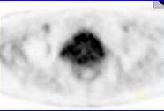
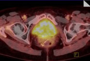
| Patient case, 67 years of age, s/p prostate carcinoma; the patient shows a rising tumor marker PSA. PET/CT performed with Gallium 68 PSMA shows several bone metastases, and several lymph nodes which are not enlarged, however, present malignant PSMA uptake. | ||
 |
 |
 |
 |
 |
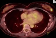 |
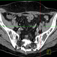
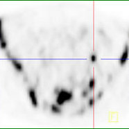
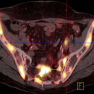
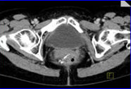
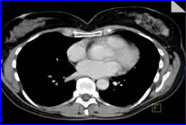
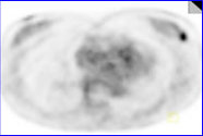
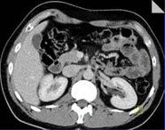
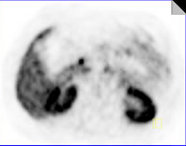
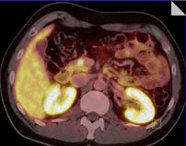
| Patient 53 years of age, s/p rectal cancer, who was transferred to FDG PET/CT after rising tumor marker CEA, with suspicion of tumor recurrence. The CT showed a suspicious soft-tissue behind the bladder without FDG-uptake in PET, so that a tumor recurrence could be excluded (top row). The whole body PET/CT images, however, showed an unexpected lesion in the left breast, which was breast cancer and the reason for the rising tumor marker (bottom row) | ||
 |
 |
 |
| Patient, 47 years of age, with recurrent hypoglycemia (low blood sugar). PET/CT performed with Gallium-68 DOTA-TATE revealed a focal tracer accumulation in the pancreas head; surgery revealed a small tumor (insulinoma) in the pancreas | ||