
PET and PET/CT for other questions
A further example for PET/CT indication is the search for inflammatory lesions which are hard to find with other diagnostic methods, or assessment of the inflammatory activity of certain diseases.
In cardiology, PET/CT is primarily used to diagnose the vitality of the heart muscle or the estimation of the scartissue, e.g. after infarction. In combination with perfusion scintigraphy, it moreover serves to assess so-called “hibernating” heart-muscle (hibernating myocardia). Further, it is used to assess the pump function and wall movement of the left ventricle e.g. prior to special pacemaker therapies.
 |
 |
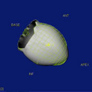 |
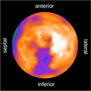
| Patient, 75 years of age, with numerous cardio-vascular risk factors and after various heart attacks. The left illustration shows an overview of the glucose metabolism in the left ventricle, the scar is localized at the right back wall (red arrows). The median image shows the corresponding three-dimensional illustration of the glucose metabolism of the left heart chamber. The right illustration shows the pump movement, calculated from the PET information, of the left ventricle; a movement disturbance of the back wall in the scar region is shown. | ||
How is the examination performed?
PET and PET/CT are performed at the hospital of the Munich university both for in-patients and out-patients and at both sites (city center and Grosshadern).
Precondition for a PET or PET/CT test is primarily finding the necessity to do this test by the treating physician, e.g. cardiologist. He will register the test within the hospital by writing a clinical order, or at the control site of the nuclear medicine for out-patients (transfer letter, doctor’s letter with exact question). Thereafter, a specialist for nuclear medicine will internally review the so-called justifying indication. If it is applicable, a date will be scheduled and given to the patient together with important rules to be complied with.
Depending on the radiopharmaceutical, the patients will have to appear on an empty stomach for the PET test with F-18 FDG (approx. 4-6 h food waiting period, whereby unsweetened tea and mineral water are allowed during this time). For PET/CT examinations, iodine containing contrast agents will be given in addition; in this respect, current blood values for TSH and creatine should be brought. Likewise, earlier results and external CT or MRT images in digital form (DVD) ought to be brought if available.
Patient having been informed and anamnesis having been taken, the course of the examination will be discussed with the patient in detail and information on possible risks of the planned administration of iodine contrast agents for computer tomography (e.g. iodine allergy) will be given. Subsequently, the blood sugar value will be measured prior to F-18 FDG tests; as a rule, it ought to be lower than 140 mg/dl. In diabetic patients, the adjustment of blood sugar values should be constant. First, after intravenous access (mostly on the back of the hand or in the crook of the arm) radiopharmaceutical will be injected into the blood stream. After application, the patient will have to wait for approx. one hour until the body has metabolized the substance or bound to the target structures. Thereafter, the actual PET and, facultatively, CT images will be performed. The scan will take between 20 and 60 minutes, depending on the question and body region to be imaged. During the entire scan, the patient will not be alone and be supported by medically technical assistants with the doctor in charge.